
Bone Is Not Solid: The Living Architecture Inside Your Skeleton

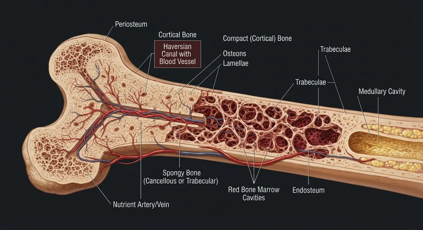
Photo: QuickAdvisor.net editorial
Key Takeaways
- Bone is living tissue threaded with blood vessels, nerves, and active cells - not a static scaffold.
- Your skeleton completely replaces most of its mass roughly every decade through continuous remodeling.
- Bone has two distinct structural zones: dense cortical bone on the outside and porous trabecular bone within.
- Mechanical stress from exercise signals bone-building cells to strengthen the skeleton.
- Bone marrow, housed inside bones, produces the majority of the body's blood cells.
The Solid Bone Myth
Most people picture bone the way they encounter it - as the dry, hard, ivory-colored material in a biology classroom skeleton. That mental image is understandable, but deeply misleading. Living bone in your body is a dynamic, vascular tissue more comparable to a busy construction site than a static scaffold.
Fresh bone is warm to the touch when exposed surgically, because it is richly supplied with blood. It bleeds when cut, aches when fractured because it carries nerves, and heals from injury because it houses cells capable of rebuilding themselves. Bone is, in every biological sense, alive.
Why Dried Bone Looks Different
Two Zones, One Structure
Look at a cross-section of a long bone - a femur, for instance - and you immediately see two distinct architectural zones working together.
The outer shell, called cortical bone (or compact bone), is dense and hard. It forms the walls of long bones and accounts for roughly 80 percent of the skeleton's total mass. Microscopic channels called Haversian canals run lengthwise through this layer, carrying blood vessels and nerves deep into the tissue.
Inside that shell lies trabecular bone (also called spongy or cancellous bone). Despite its name, this isn't weak - it's a three-dimensional lattice of mineralized struts arranged to handle compressive forces efficiently, much like the internal bracing of a modern bridge. The spaces between those struts are filled with bone marrow.
This combination of dense exterior and porous interior gives bone an extraordinary strength-to-weight ratio - strong enough to bear the body's full load, light enough to allow movement.
~80%
Skeleton mass that is cortical bone
Cortical bone forms the dense outer walls of most bones and constitutes the majority of total skeletal mass, according to standard anatomical references.
206
Bones in the adult human body
Newborns have closer to 270 bones; many fuse during childhood and adolescence, resulting in the adult count of 206.
~2 million
Red blood cells produced per second
Bone marrow is responsible for continuously generating red blood cells at this remarkable rate to replace those that naturally age out of circulation, according to hematology literature.
The Cells That Run the Construction Site
Bone's living character comes from three principal cell types operating in coordinated cycles:
- Osteoblasts build new bone by secreting collagen proteins that form a flexible scaffold, which then hardens as calcium phosphate minerals crystallize within it.
- Osteoclasts dissolve old or damaged bone tissue by releasing acids and enzymes - a process called resorption. This isn't destruction for its own sake; it removes microscopic cracks before they propagate and returns calcium to the bloodstream where the body needs it.
- Osteocytes are former osteoblasts that became embedded in the hardened matrix. They act as sensors, detecting mechanical strain and signaling the body to increase or decrease remodeling activity accordingly.
The balance between osteoblast and osteoclast activity determines whether bone mass stays stable, increases, or decreases over time. Exercise tips the balance toward building; prolonged immobility or certain hormonal changes tip it toward loss.
Why This Biology Matters for Everyday Health
Understanding bone as living tissue changes how you interpret common health advice. The push to stay physically active, eat adequate calcium and vitamin D, and avoid prolonged sedentary stretches isn't arbitrary - it directly targets the cellular machinery that keeps remodeling balanced.
Weight-bearing exercise loads the skeleton, which osteocytes detect and interpret as a signal to reinforce the structure. Calcium provides the mineral raw material osteoblasts incorporate into new tissue. Vitamin D is essential for absorbing that calcium from food in the digestive system. These aren't isolated tips; they're inputs to the same biological system.
This is general health information, not personal medical advice. Anyone concerned about bone density or skeletal health should speak with a qualified healthcare provider, who can assess individual risk factors and appropriate next steps.
This article is for informational purposes only and does not constitute medical advice. Consult a qualified healthcare professional for any concerns about your bone health or overall wellbeing.
Frequently Asked Questions
The content on this site is for informational purposes only and is not a substitute for professional advice. Always consult a qualified professional for guidance specific to your situation.